Neonatal bilirubin is a yellow breakdown product of normal heme catabolism in newborns. Its levels are elevated in certain diseases and responsible for the yellow color of bruises.
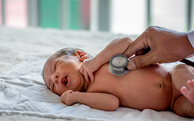
Risk to Newborns
Hyperbilirubinemia is one of the most common problems encountered in term newborns. Up to 60% of term newborns have clinical jaundice in the first week of life.
High Bilirubin Levels are Common in Newborns
High bilirubin levels in newborns are relatively common, especially within three days of birth. This is sometimes called physiologic jaundice of the newborn. Within the first 24 hours of life, up to 50% of full-term newborns, and an even greater percentage of pre-term babies, may have a high bilirubin level. After birth, newborns begin breaking down the excess red blood cells (RBCs) they are born with and, since the newborn’s liver is not fully mature, it is unable to process the extra bilirubin, causing levels to rise in the blood and other body tissues. This situation usually resolves itself within a few days.
Principle of Operation
Total Bilirubin – The maximum light absorption of bilirubin occurs at a wavelength of 454 nm. Oxyhemoglobin (HbO2) has nearly equal absorbance at 454 and 540 nm. In the BR2, the absorbance at 540 nm (HbO2) is subtracted from the absorbance at 454 nm (TBR + HbO2). The remainder corresponds to TBR concentration corrected for oxyhemoglobin up to 600 mg/dL. Light from an incandescent source is collimated and directed through a cuvette containing the diluted sample. A fiber optic beam splitter with 454-nm and 540-nm channels splits the transmitted light, each made monochromatic by a narrow-bandpass interference filter. Matched solid-state photo-detectors and signal processing electronics measure the transmittances at 454 and 540 nm and display the TBR concentration digitally.
Direct Bilirubin – Direct Bilirubin is measured as the change in absorbance at 540 nm due to the formation of azobilirubin, using a two-minute, timed-endpoint modification to the Malloy and Evelyn method. After TBR has been measured on a sample, that same sample is acidified with HCl, establishing an absorbance baseline. Next, diazotized sulfanilic acid is added. Under these conditions, the conjugated (Direct) bilirubin fraction will react to form azobilirubin, which has maximum light absorption at 540 nm. At the end of the two-minute reaction period, the digital display locks in to hold the DBR readout until the next test is initiated.